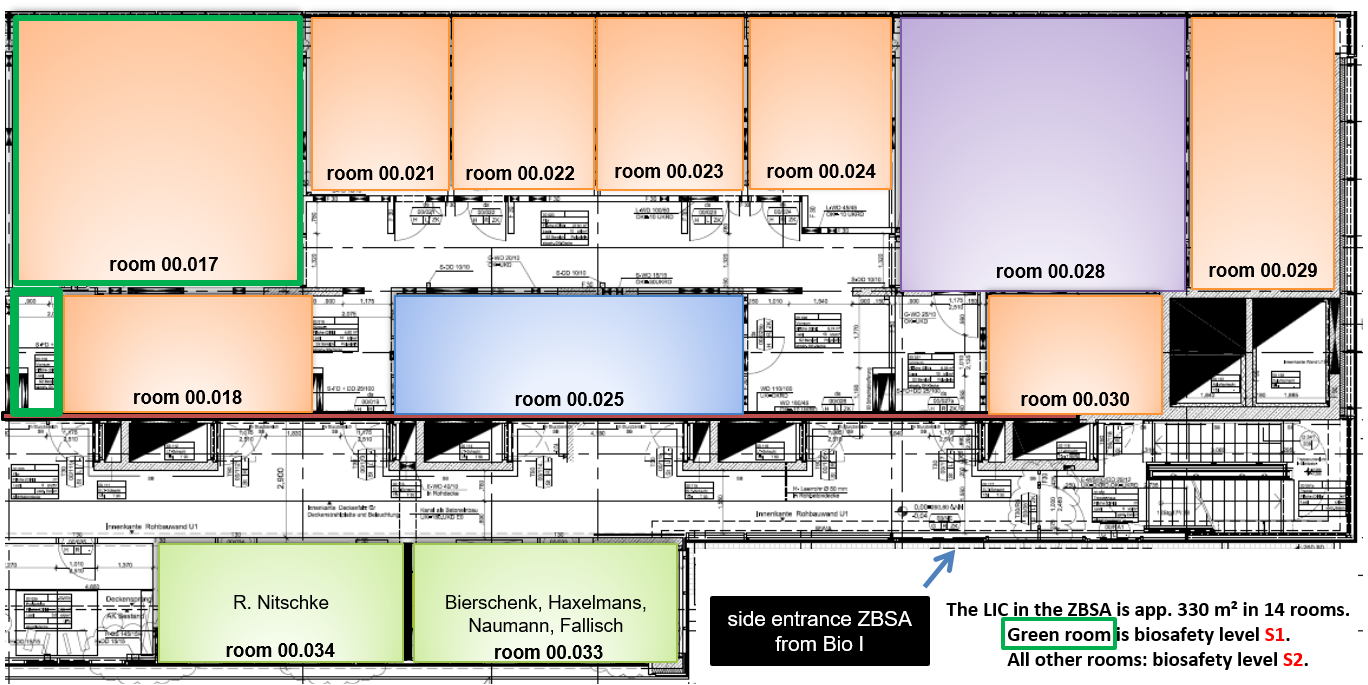
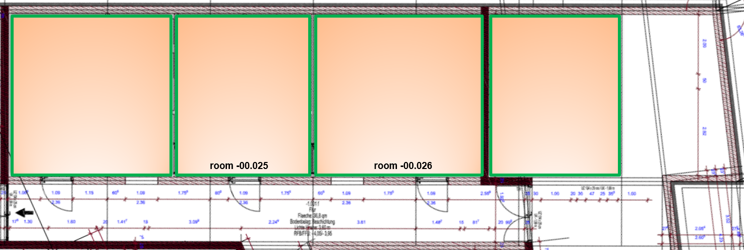
The Life Imaging Center (LIC) is a central core facility of the Albert-Ludwigs-University Freiburg open to members of all Faculties and the Universitätsklinikum. Currently around 250 active users from six Faculties (Biology, Medicine, Chemistry and Pharmacy, Environment and Natural Resources, Mathematics and Physics, and Engineering) use the LIC infrastructure. LIC users come from more than 68 workgroups and the combined usage time of the LIC microscopes in 2019 was about 10.450 hours. The LIC has approximately 60 – 80 new users per year and approximately the same number is leaving the facility each year. Users from external institutions (e.g. Max Planck Institutes) and from national and international universities (e.g. Basel, Strasbourg, Haute-Alsace) are able to use the LIC infrastructure in collaboration projects. More details about the application procedure and facility guidelines can be found at Application & Booking. You can find contact information for the head and staff of the LIC facility at Team website. The LIC consists of 14 rooms in the Hilde-Mangold Haus (HMH) with approximately 400 m² (9 microscopy rooms with 20 microscope set-ups (3D-STED, confocal FLIM, two 2-photon confocal microscopes, 4 conventional confocal microscopes with super-resolution (Zeiss Airyscan or Leica Lightning), lightsheet microscope, 3 high-end widefield ratio-imaging set-ups incl. TIRF, 4 fluorescence stereo microscopes, 3 special set-ups for screening). The LIC microscopes are hosted in Biosafety level S2 and S1 environments, connected to a large wet lab area and a cell culture room, both Biosafety level S2 classified. Additionally, the LIC offers a large computer lab with 12 high-end workstations for image processing and analysis. The LIC also has three more lab rooms with 4 confocal microscopes in the Signalhaus, Schänzlestr. 18 and two additional labs with associated equipment, hosting 3 LSM systems in a neighboring building of the Signalling Campus Freiburg (SCF).
Techniques and Samples:
Most modern microscopic techniques (2-Photon, FLIM, FRET, FRAP, FLIP, FLAP, photo-conversion, photo-activation, uncaging, spectral unmixing, multi-position and large area recording, TIRF, HILO microscopy, structured illumination, microinjection) can be performed in combination with live cells or organisms on the available microscope set-ups. The LIC has a major focus on live cell imaging of all kind of cells, organisms and culture systems used in cell signaling research, developmental biology and neurobiology. Samples are: Embryos (Danio rerio, Drosophila melanogaster, Mus musculus, Xenopus laevis), model organisms or cell clusters (C.elegans, spheres, cysts), plants (root, leave, growth cone), organoids, neuronal cultures, explants and biopsies from tissue slices, spheroids, flow cultures as well as cells cultured on glass slides. LIC is specialized in long time observation and experiments on these objects, high resolution large area and/or multiple location recordings of thick objects with 2P and UV/VIS excitation. A further specialty is culturing and permanent microscopic observation of cells and spheroids over periods of up to 10 days (also under flow conditions in special chambers). All types of fluorescent proteins including many photo-activatable and photo-convertible are used. We have a large library of fluorescent proteins (more than 150) available for our users via the Signalling Factory and we perform test experiments with these constructs before giving them out to our customers. See also: Available excitation wavelengths for imaging at LIC (PDF).