LIC IT hardware infrastructure
Computer lab for data analysis (ground floor room 00.041)
Computer lab overview (PDF) (where to find the dedicated workstations)
- 12 high-end PCs Workstations with up to 24 cores, each 128-512 GB RAM, Nvidia GPUs with up to 24 GB VRAM, local storage capacity each between 4-40 TB (total local capacity 250 TB in computer lab), 30-38″ TFT monitors (2560 x 1600, 3840 x 2160 or 5120 x 2160). All systems are fully equipped for offline analysis including local deconvolution options, for installed software details (PDF).
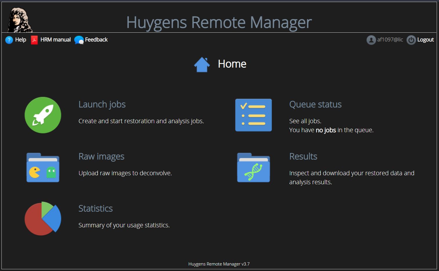
- Huygens Core server (HRM) for deconvolution (256 GB RAM, 2 CPUs, 28 cores, 24 GB GPU VRAM, 6 GB SSD RAID 5, 28 TB storage RAID 5) can be accessed by http://slurms.lic.ads.zbsa.privat/hrm (only accessible within the network of Freiburg University), Licenses available: deconvolution for confocal, multiphoton, spinning disk, STED, light-sheet, widefield, Airyscan (ZEISS), colocalization analysis, light-sheet fusion, object stabilizer, timeseries. See our manual section for detailed instructions on how to use the Huygens server.
- Specialized high-speed large capacity data storage of 160 TB for light sheet data storage.
- NextCloud to send large amounts of data (for more information contact the LIC team (lic@imaging.uni-freiburg.de).
Software Infrastructure
Software for image analysis
Software overview (PDF) (where to find our software tools)
Software
Category
Example task
Available on computer
User Interface
Presentation
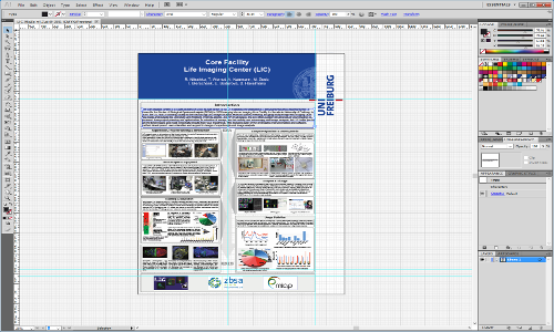
Preparation of vector graphics
Preparation of scientific posters
001, 003, 004, 010, 011, 012
Editing
Presentation
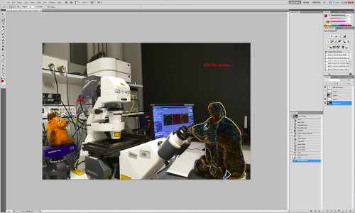
Extensive image editing tools, pixel graphics
001, 003, 004, 006, 011, 012
3D/4D visualization
Image analysis
3D visualization
3D analsis
Time-lapse (4D), movie
009, Light-Sheet-Ana
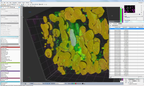
Image analysis of Biostation images
setup-specific data editing / analysis
all except Light-Sheet Ana
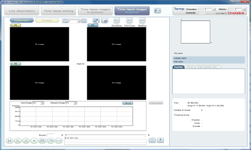
Image analysis
Editing
Counting and measuring
Object tracking
co-localization
Many Plug-Ins (open source)
all
Post-Processing / Deconvolution
Deconvolution
all (floating, limited number of licenses)
Post-Processing / Deconvolution
Deconvolution
all (floating, limited number of licenses)
3D/4D visualization
Image analysis
Surface reconstruction
Tracking
Measurement
all (floating, limited number of licenses)
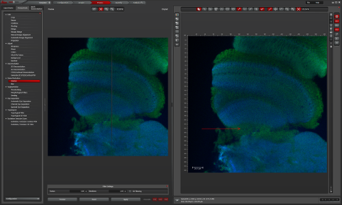
Leica Application Suite
(LAS-AF) for TCS SP8
Image analysis of Leica images
setup-specific data editing / analysis
004, 005, 007, 009
Data analysis
Presentation
Plotting of fuctions and data
Implementation of algorithms
all (runtime)
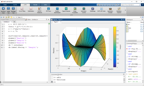
Image analysis
setup-specific data editing / analysis
Fluorescence ratio imaging
004, 008, 009
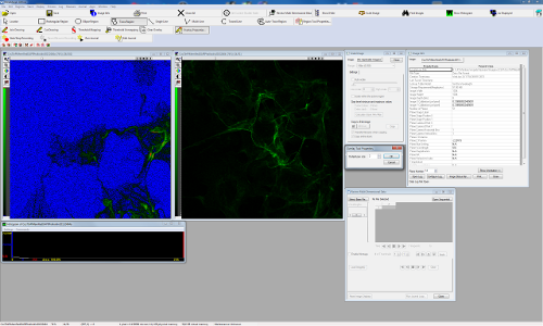
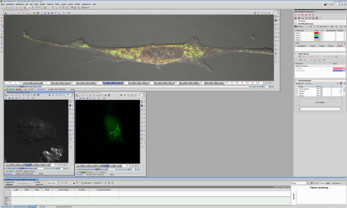
Image analysis of Nikon images
setup-specific data editing / analysis
all except Light-Sheet Ana
3D image analysis
Surface reconstruction
Tracking
Measurement
all except Light-Sheet Ana
(no maintenance)
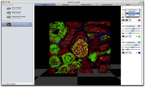
Software
Category
Presentation
Example task
Preparation of vector graphics
Preparation of scientific posters
Available on computer
001, 003, 004, 010, 011, 012
User Interface

Software
Category
Editing
Presentation
Example task
Extensive image editing tools, pixel graphics
Available on computer
001, 003, 004, 006, 011, 012
User Interface

Software
Category
3D/4D visualization
Image analysis
Example task
3D visualization
3D analysis
Time-lapse (4D), movie
Available on computer
009
User Interface
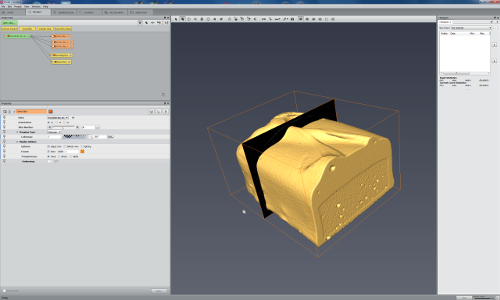
Software
Category
3D/4D visualization
Image analysis
Example task
3D visualization
3D analsis
Time-lapse (4D), movie
Available on computer
009, Light-Sheet-Ana
User Interface

User Interface
Software
Category
Image analysis of Biostation images
Example task
setup-specific data editing / analysis
Available on computer
all except Light-Sheet Ana
User Interface

Software
Category
Image analysis
Editing
Example task
Counting and measuring
Object tracking
co-localization
Many Plug-Ins (open source)
Available on computer
all
User Interface

Software
Category
Image analysis
Editing
Presentatio
Example task
2D/3D analysis
graphing and plotting
Available on computer
none
User Interface
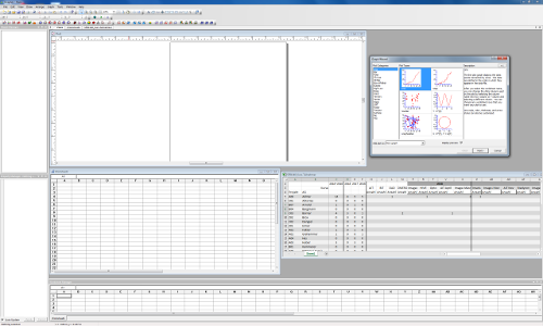
Software
Category
Post-Processing / Deconvolution
Example task
Deconvolution
Available on computer
all (floating, limited number of licenses)
User Interface

Software
Category
Post-Processing / Deconvolution
Example task
Deconvolution
Available on computer
all (floating, limited number of licenses)
User Interface

Software
Category
3D/4D visualization
Image analysis
Example task
Surface reconstruction
Tracking
Measurement
Available on computer
all (floating, limited number of licenses)
User Interface

Software
Category
Editing
Example task
Viewing all kinds of formats
Simple editing tasks
Available on computer
all
User Interface
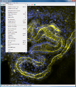
Software
Leica Application Suite
(LAS-AF) for TCS SP8
Category
Image analysis of Leica images
Example task
setup-specific data editing / analysis
Available on computer
004, 005, 007, 009
User Interface

Software
Category
Data analysis
Presentation
Example task
Plotting of fuctions and data
Implementation of algorithms
Available on computer
all (runtime)
User Interface

Software
Category
Image analysis
Example task
setup-specific data editing / analysis
Fluorescence ratio imaging
Available on computer
004, 008, 009
User Interface

Software
Category
Presentation
Example task
Writing documents
Preparing presentations
Managing numerical data
Available on computer
all
User Interface
Software
Category
Image analysis of Nikon images
Example task
setup-specific data editing / analysis
Available on computer
all except Light-Sheet Ana
User Interface

Software
Category
Data analysis
Presentation
Example task
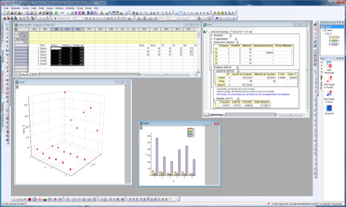
Scientific graphing and plotting
Available on computer
all except Light-Sheet Ana
User Interface
Software
Category
Screen capture
presentation
Example task
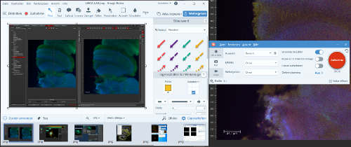
Video creation for presentation
Available on computer
all except 005
User Interface
Category
Video preparation
Presentation
Example task
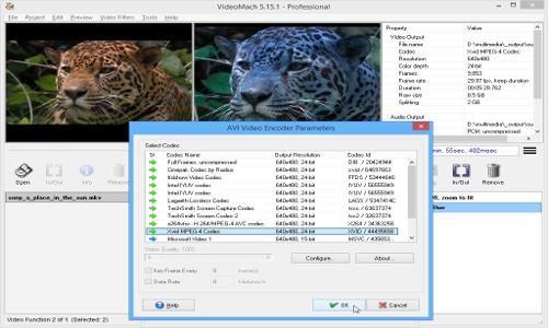
Video creation for presentation
Available on computer
all except Light-Sheet Ana
User Interface
Software
Category
3D image analysis
Example task
Surface reconstruction
Tracking
Measurement
Available on computer
all except Light-Sheet Ana
(no maintenance)
User Interface

Software
Category
Image analysis of Zeiss images
Example task
setup-specific data editing / analysis
Available on computer
all except 005
User Interface
Software
Category
Image analysis of Zeiss images
Example task
setup-specific data editing / analysis
Available on computer
all expect 011
User Interface
Macro
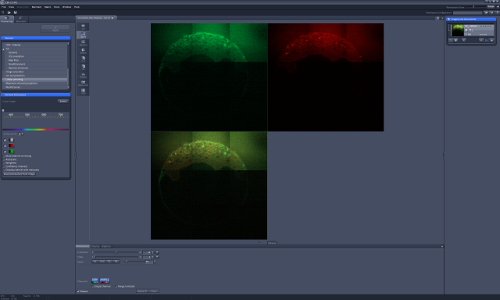
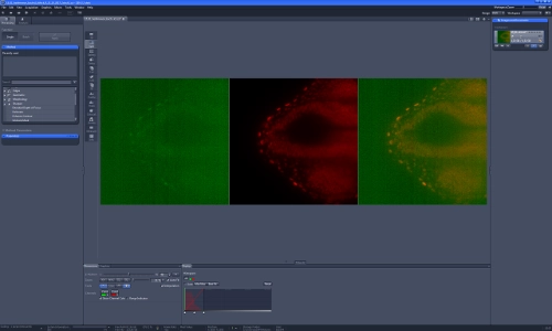
Over the last 4 years the LIC team has developed a specialized software add-on for all ZEISS LSM (family 5 and 7 instruments) running the ZEISS ZEN black software (fully compatible with ZEN 2009 and 2010 on 32bit WinXP and Win7). Support for 64 bit Win7 and ZEN 2011 and 2012 will be available soon.
The software allows half-automatic and automatic image recording of multiple 2D or 3D areas over time using different scan settings adapted to the specimen. Recognition of image structures, properties or dynamic changes by the software or outside image analysis tools can be combined with the software to select or exclude, correct or trace the recording areas without or with user interaction. Automated image recording based on guidelines defined by the user for the specific experiment is achieved.
The following features are currently implemented:.
1.) Automated large area focus position correction based on cover-glass tilt
2.) Measurement of objective xy-axis shift with position correction after objective change
3.) Multi-resolution Overview image recording with an image based coordinate system
4.) Synchronisation of sample position and overlay of newly recorded confocal images to overview images recorded on any microscope or imaging device (for multimodal imaging with different devices or repetitive, non-continuous recordings of time series or large samples)
5.) Multi-position and/or multi-tile recordings also based on dynamic event recognition with independent or linked recording conditions and time schemes for each tile position. Location and tile definitions manually by drawing on the overview or automated based on sample properties
6.) Heterogeneous spectral (lambda), multi-track recordings over time
7.) Self adjusting 3D-stack position and dimension based on user definable properties
8.) Online multi-object tracing over time in 3D based on image properties
9.) Data processing module for image retrieval, export and further processing
The software is tested and used in a large beta-test at 12 European imaging facilities and research institutes. The software is currently used for large area recordings of whole chicken embryos, large brain slices; multi-point/multi-tile 2D/3D time-lapse and tracing of Xenopus embryos, cultured cells, and growing Arabidopsis roots; as well as control of external devices (lasers) for photo-manipulation based on image content. The LIC Macro was presented with talks and posters at “Focus on Microscopy” (FOM) 2010, 2012 and 2013 and ELMI meeting 2012. Short Manual “How use LIC Macro” (LIC). Outside institutes interested in the usage of the software please contact the LIC team under: lic@imaging.uni-freiburg.de
Quality Management
In response to both users’ and customers’ demands, LIC has developed an internal Quality Management to ensure high quality of facility infrastructure and services. One of the main objectives of this quality management is the consistent enhancement of facility overall performance to provide always access to state of the art technology in the field of life science.
A quality assurance system for the technical equipment in the LIC was established stepwise over the last years. We also collect all input from our users about quality control, new techniques, hardware, and software in a database and discuss this in our weekly LIC staff meetings and as well as in budget planning rounds with local funders from the University and the Faculties.
The following tests and maintenance work as well as supporting measures are carried out on regular basis (see further information: quality management manual ).
Daily:
- Visual inspection and necessary cleaning of all important parts and accessories of the microscope set-ups (objectives, motorized tables, sample holders, table inserts, immersion fluids and applicators, completeness of cleaning equipment for objectives, microscope incubators, check aqua stop)
- Cell culture room: Check water bathes and incubators
- Temperature of incubators, deep freezers, freezers and refrigerators are permanently monitored and alarm is sent out by mobile phone in case of error
Weekly:
- Check the storage capacity of the local hard drives including the OS hard drive
- Remove all temporary files
- Backup of the OS hard drive
- Check, if objective protection works properly
- Inspection of the objectives under a stereo-microscope, Control images are taken under the stereo microscope from each front lens to monitor damages like scratches, Cleaning with a special lens cleaning fluid from Zeiss under permanent rotation of the lens to prevent drying of dirt on the front lens surface
- Ponit-Spread-Function (PSF) measurement, if lens has new scratches or other problems
Monthly:
- Delete expired local user accounts
- Check the pinhole and collimator alignment with a test sample for UV, VIS and 2-photon using a standard test procedure. We have a system set-up, which automatically resets the correct pinhole settings after each session, so that any misalignment done by a user will be corrected the next time the confocal software is restarted. So this assures that only the LIC staff can change pinhole and collimator settings after performing an exact calibration procedure.
- Confocal scanner calibration with a special software tool to keep the imaging performance high
- Shading problems are evaluated with color slides from Chroma
- Laser out-put power is documented for all laser lines with a special power-meter directly mounted on the microscope table
- Out-put power of the fluorescence illumination on widefield microscopes is documented with a special power-meter directly mounted on the microscope table
Quality control
(covering monitoring of user satisfaction and project success in terms of published results):
- We run a trouble-ticket system, where our users can, with a click on an icon on the desktop, give us directly input including screenshots or attach files.
- We have run a user satisfaction survey using SurveyMonkey up to now only once in 2010, but will repeat this every half year in the future.
- We try to follow up the success of projects by actively writing to all project leaders once per year to send us the PDF of the papers, they published with images/results enabled by the LIC infrastructure. However, these kind of requests have usually only a relative poor feedback rate of 50%, so we will soon start with a special kind of bonus systems for users, who acknowledge the work of the LIC in their papers. We have included this in our Usage Conditions and Rules for the LIC
for further question send E-mail to : lic@imaging.uni-freiburg.de
VIBE-Z
The LIC has actively taken part in the development and realization of ViBE-Z project. ViBE-Z includes some specialties like stitching, two-sided sample recording followed by image-fusion and absorption correction, which often greatly enhance imaging quality and visualization of large, thick objects.
ViBE-Z developed originally for zebrafish data, contains currently a database with precisely aligned gene expression patterns (1?m resolution), an anatomical atlas, and a software. This software creates high-quality data sets by fusing multiple confocal microscopic image stacks, and aligns these data sets to the standard larva. A detailed description of the ViBE-Z framework and its applications is provided in Ronneberger et al. (2012).
References:
Olaf Ronneberger, Kun Liu, Meta Rath, Dominik Rueß, Thomas Mueller, Henrik Skibbe, Benjamin Drayer, Thorsten Schmidt, Alida Filippi, Roland Nitschke, Thomas Brox, Hans Burkhardt, and Wolfgang Driever: ViBE-Z: A Framework for 3D Virtual Colocalization Analysis in Zebrafish Larval Brains. Nature Methods 9(7) pp. 735-742 (2012) (doi:10.1038/nmeth.2076). (Supplementary Movies)
Meta Rath, Roland Nitschke, Alida Filippi, Olaf Ronneberger, and Wolfgang Driever:Generation of high quality multi-view confocal 3D datasets of zebrafish larval brains suitable for analysis using Virtual Brain Explorer (ViBE-Z) software. Nature Protocol Exchange 22/06/2012 doi:10.1038/protex.2012.031


