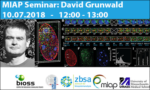
Historically, major advances in biology have rapidly followed major advances in microscopy, often driven by biologists’ desires to visualize ever and ever smaller objects. Here I present advances in studying the real-life dynamics of molecules in the cell nucleus using RNA aptamers, CRISPR based DNA labeling technology, 2D and simultaneous 3D single molecule real time microscopy in to bypass the limits of diffraction. Although technological advances have been made, issues like unspecific background, limited signal, and optical aberrations still limit our imaging abilities, but smart image-processing and –analysis strategies for quantification can help to further push the limits. However, to increase data quality and fidelity for biological measurements in images requires microscope and experiment specific meta-data for the characterization of data obtained through fluorescence microscopy. I will present tools and meta data standards currently developed as part of NIH’s 4D nucleome consortium to address these needs.
Albert-Ludwigs University Freiburg
Microscopy and Image Analysis Platform (MIAP)
Life Imaging Center (LIC)
Center for Biological Systems Analysis (ZBSA)
Room -01.026
Habsburgerstr. 49
79104 Freiburg im Breisgau
David Grunwald, PhD
University of Massachusetts Medical School
368 Plantation Street, AS4.1043
Worcester MA 01605
Phone: 774-455-3632
E-Mail: David.Grunwald@umassmed.edu
MIAP
web: https://miap.eu
e-mail: info@miap.eu
BIOSS
web: http://www.bioss.uni-freiburg.de/
e-mail: kontakt@bioss.uni-freiburg.de
A joint Network for scientific Imaging and Image Analysis Infrastructure
Phone: +49 761 203 2934
Email: info@miap.eu
Contact Form